
Digital Radiography
AMHERST VILLAGE DENTAL
DIGITAL RADIOGRAPHY
X-rays are a crucial aspect of the medical world. X-rays show both doctors and patients interior images of the patients’ bodies, allowing for an effective diagnosis. X-rays are also used in the dental world to diagnose and treat oral health problems such as developing cavities or infected wisdom teeth. At Amherst Village Dental, our team can effectively treat a variety of oral health problems. There are two common ways to take x-ray photos: film x-ray and digital radiography.
Digital Radiography Has Become An Important Tool In Dentistry
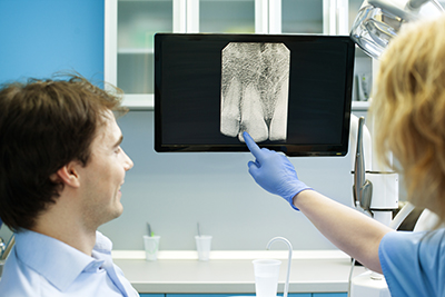
Digital radiography is a modern approach for taking x-ray images. Film radiography, like an analog camera, has been used for many years and is still used to this day. However, dental offices all over the country have switched to digital radiography. Digital radiography is a powerful and versatile means of taking images. After the picture is taken, the picture is instantly available to view. Digital radiography also emits less radiation than film x-ray machines. Furthermore, digital radiography photos come out larger which enables our professionals to analyze issues more effectively.
Intraoral X-Rays
Intraoral x-rays are pictures taken inside of the mouth. Bitewing x-rays require the patient to bite down on film. Bitewing x-rays show the details of upper and lower teeth in one area of the mouth. The area that gets pictured covers the area from the crown to the supporting bone. Bitewing x-rays are used to assess damage from tooth decay and gum disease, as well as to monitor dental devices such as crowns and fillings. Periapical x-rays have a similar area of coverage as bitewing x-rays, but they detect abnormalities in root structure or bone structure, making them essential for treating periodontitis and abscesses.
Extraoral X-Rays
Panoramic x-rays show all of a patient’s teeth in a single image. Panoramic x-rays help with the planning of dental implants, the detection of impacted wisdom teeth, and the detection of malignant cysts. Multi-slice computer tomography focuses on a single layer of the mouth and is used to examine structures that would otherwise be difficult to focus on. Cephalometric projections take a picture of the patient’s entire head, and these pictures assist our team in developing the right treatment plan for the patient. Sialography is a process in which the patient has dye injected into their salivary glands, which makes the glands visible in an x-ray. Sialography detects problems in the salivary glands which might lead to oral health issues such as dry mouth and gum disease. Cone beam computerized tomography takes a three-dimensional picture of the patient’s entire body. Our team uses cone beam computerized tomography to identify tumors and fractures and to prepare for tooth extractions and installing dental implants.
Give Us A Call If You Require Oral Care
Don’t let failing and missing teeth keep you from feeling confident or from doing the things you love. Call us today at 603-673-5510 to invest into your health, your life, and your future.